

EyeTX Vision Center
Our board-certified eye doctors provide the highest quality of care performing everything from routine eye exams to diagnosing, treating, and managing eye diseases.
Find an Eye Doctor Near You
About EyeTX Vision Center
Founded in San Antonio, EyeTx Vision Centers provide quality vision solutions for many Texas communities. EyeTx Optometrists are Therapeutic & Glaucoma certified. They specialize in routine eye care, contact lens, gas permeable fittings, bi-focal contact lens fitting, Kerotoconus fittings, and Ortho-K fittings. We offer Gucci, Oakley, RayBan, Michael Kors, and more! Most insurances are accepted. Call or schedule your eye exam online.

Find an Eye Doctor Near You
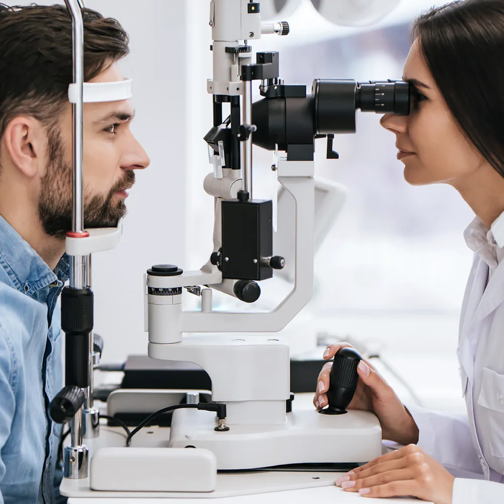
At EyeTX Vision Center we believe in using the latest technologies to aid us in doing everything comprehensively—right down to your basic eye exam.